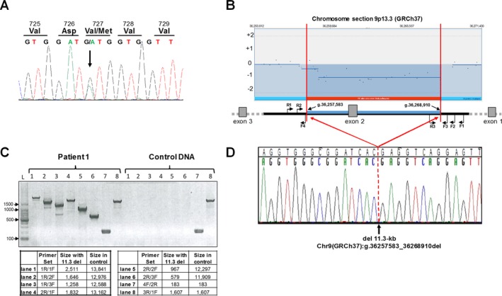
Figure 2.

GNE mutation analysis. (A) Sanger sequencing of GNE exon 13 confirmed the heterozygous GNE mutation [NM_001128227.2:c.2179G>A;p.Val727Met] in both siblings (Patient 1 displayed). (B) Enlarged region of chromosome 9 (GRCh37) aCGH analysis that showed heterozygosity of a large (>10 kb) region in both patients. Primer locations for deletion analysis are indicated (not to scale, note that GNE gene is transcribed on reverse strand). (C) The size and breakpoints of the deletion were established by PCR analysis across the deletion; expected fragment sizes for each primer set are indicated. (D) Sanger sequencing across the deletion (Primer 1R; reverse sequence shown) determined the exact breakpoints and deletion size as: Chr9(GRCh37):g.36257583_36268910del (del 11,328‐bp).
