
Figure 6.
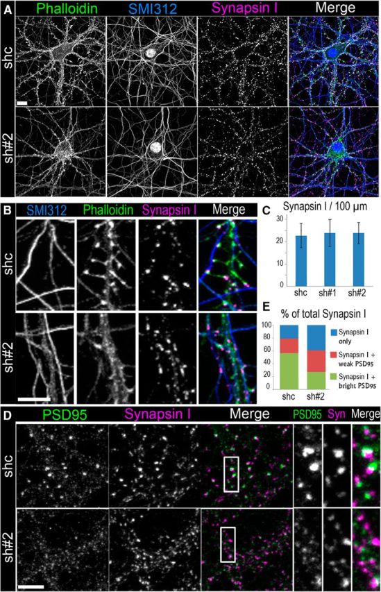
Depletion of βIII spectrin results in formation of aberrant synapses. A, B, Fluorescence staining of 17 DIV neurons transfected with control shRNA (shc) or βIII spectrin shRNA (sh#2) with phalloidin, axonal marker SMI312, and synapsin I antibody. Scale bar, 10 μm. B, High-magnification images showing spiny synapses in a control cell (top) and shaft synapses in a βIII spectrin-depleted cell (bottom). Scale bar, 5 μm. C, Average numbers of synapsin I puncta per 100 μm of axon length (shc: N = 664 puncta in 9 cells; sh#1: N = 631puncta in 10 cells; sh#2: N = 615 puncta in 10 cells). Error bars indicate SD. D, Staining of PSD95 and synapsin I in control (shc) and βIII spectrin-depleted (sh#2) cells. Scale bar, 5 μm. Boxed areas are zoomed at right. E, Quantification of synapsin I puncta associated with “bright” or “weak” PSD95 puncta or not associating with PSD95 puncta (“synapsin I only”) in control and βIII spectrin-depleted cells (shc, N = 694 puncta in 5 cells; sh#2, N = 578 puncta in 5 cells).