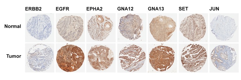
Figure 3. IHC analysis for the expression of target proteins in the endometrial cancer TMA.
A TMA, constructed from the patient FPPE blocks of 1 mm diameter cores was subjected to IHC analysis using antibodies to specific proteins encoded by the representative target genes for the putative tumor suppressor miRs as detailed in the text. Sample micrographs of the TMA spots developed with 3, 3’-diaminobenzidine (DAB)/HRP staining (brown) for the respective protein are presented; magnification: X 10).

