
Figure 3.
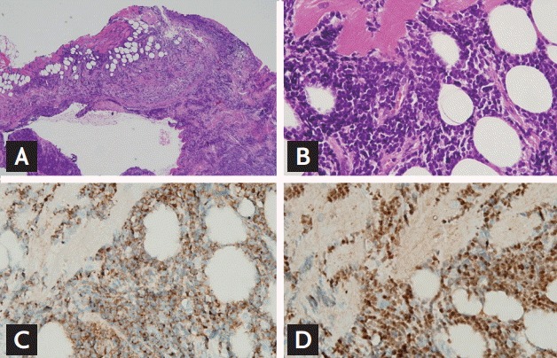
Recurrent acute lymphoblastic leukemia (ALL) in the heart. (A) Ventricular biopsy showing diffuse infiltration of monotonous small cells in the myocardium and pericardium (H&E, ×40). (B) Lymphocytes showing infiltration and encroachment on cardiomyocytes. The atypical lymphocytes are medium-sized with round to oval nuclei showing vesicular chromatin and inconspicuous nucleoli, and have a scanty cytoplasm (high nuclear/cytoplasmic ratio; H&E, ×400). (C) Lymphocyte membranes expressing the pan-B-cell marker CD22 (×400). (D) Presence of terminal deoxynucleotidyl transferase in the nuclei of lymphocytes indicating their lymphoblastic nature (×400).