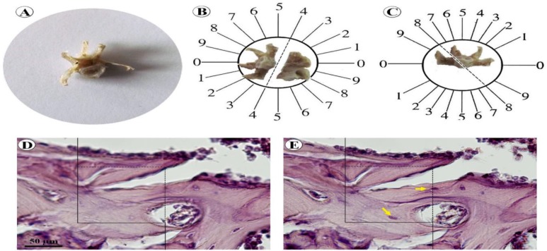
Figure 1.
The stereological methods. A lumbar vertebra was dissected out (A). Orientator method was used to obtain isotropic uniform random sections of the bone (B, C).Optical dissector method (D). An unbiased counting frame was lied on the images (E). The cell nuclei, which were not in contact with the forbidden lines (bold lines), were counted (arrows

