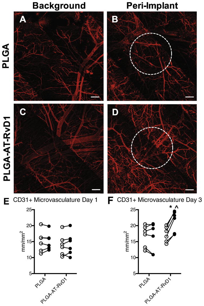
Fig. 8.

AT-RvD1 delivery enhances growth of CD31+ microvasculature. Whole mount confocal images of dorsal tissue at day 3 following treatment with AT-RvD1-loaded PLGA films. (A, C) Background microvasculature and peri-implant vasculature treated with (B) PLGA films and (D) AT-RvD1-loaded films. Quantification of microvascular length density at (E) day 1 and (F) day 3 after film implantation. ○ = background vasculature, ● = peri-implant vasculature. Statistical analysis was performed using Repeated Measures two-way ANOVA with Sidak's post-hoc test for multiple comparisons *p < 0.05 compared to background, ˆp < 0.05 compared to PLGA implant, n = 6, lines connect paired analysis of background and peri-implant vasculature in each animal. Scale bars, 100 μm.
