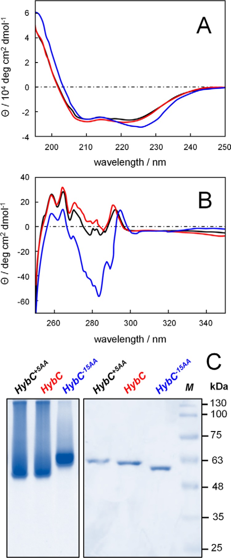
Figure 6.

Conformational changes of HybC upon removal of the C-terminal peptide. A, far-UV CD spectra; B, near-UV CD spectra of HybC+5AA (black), HybC (red), and HybC−15AA (blue). The five-amino acid extension on HybC does not lead to significant conformational changes. In contrast, removal of the C-terminal extension induces clear conformational changes in secondary and tertiary structure (compare red and black versus blue). C, native PAGE analysis (left) shows clear changes in migration behavior after removal of the C-terminal extension. The HybC−15AA protein band is sharper and migrates slower than HybC and HybC+5AA variants, indicative of a different conformation. SDS-PAGE (right) reveals the only small differences in mass between HybC variants.
