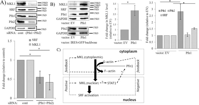
Figure 7.
Initial evidence of sensitivity of MKL and SRF expression to perturbations of Pfn. A, immunoblot analyses of MDA-231 extracts showing the effect of co-depletion of Pfn isoforms (via transfection of pooled siRNAs targeting Pfn1 and Pfn2) on MKL1 and SRF levels. The bar graph summarizes the quantification (mean ± S.D.) of the fold-changes of MKL and SRF (n = 3 experiments; *, p < 0.05). B, immunoblot analyses of MDA-231 extracts showing the effect of transient overexpression of Pfn1 (cloned into GFP-IRES backbone vector) on MKL1, SRF, and Pfn2 levels (cells transfected with the GFP-IRES backbone vector served as a control group). The bar graphs show the mean ± S.D. values of the fold-changes in MKL1, SRF, and Pfn2 expressions with respect to the corresponding control transfection condition (n = 3 experiments; *, p < 0.05). GAPDH blots serve as the loading control. EV, empty vector. C, hypothetical model of actin/MKL/Pfn/SRF signaling circuit. This model integrates the current findings of Pfn being regulated downstream of MKL in an SRF-independent manner through STAT and our initial evidence supporting Pfn's ability to also modulate MKL and in turn SRF expression, thus possibly enabling a positive feedback loop. SRF activation can either elicit a feedforward (through promoting actin polymerization, MKL expression) action amplifying the response or a negative feedback action (through elevating G-actin level) thus dampening the response beyond a certain limit (dashed lines with question marks indicates mechanisms unknown).

