Figure 5.
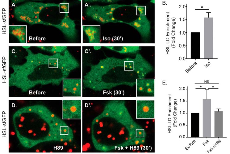
HSL is recruited to hepatocyte LDs in response to cAMP agonists. A and B, fluorescent images (A) of Hep3B cells expressing HSL-sfGFP and labeled with the LD marker MDH (red) show an increase in LD-localized sfGFP intensity following isoproterenol treatment (50 μm, 30 min), which is quantified in B (n = 13 cells from three independent experiments). *, p < 0.05. C–E, fluorescent images and the corresponding graph show treatment with forskolin also causes HSL recruitment to the LD, a response that is blocked by pretreatment with 10 μm H89 (n = 13–19 cells from 3 independent experiments). *, p < 0.05. Statistical analysis of fold change was done using a two-tailed paired t test. *, p < 0.05.

