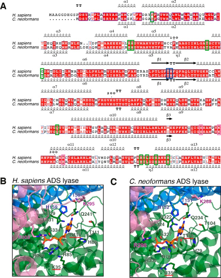
Figure 6.
Active site comparison of human and C. neoformans ADS lyases. A, sequence alignment of ADS lyases from human and C. neoformans. Residues corresponding to the active site are boxed represented by colors corresponding to subunit A (green), B (blue), and C (pink). B and C, active-site residues from human (His-86, Arg-85, Thr-111, Gln-241, Arg-329, Leu-331, Arg-338, His-159, Arg-303, Lys-295, Thr-158) and Gly-116 and Lys-35 underlined in red (B) and C. neoformans (His-79, Arg-78, Thr-104, Gln-234, Arg-322, Leu-324, Arg-331, His-152, Arg-3296, Lys-288, Thr-151) and Thr-118 and Arg-35 underlined in red (C) ADS lyases, with side chains of active-site residues shown in stick representation. The colors correspond to the subunit in which they belong. The bound AMP (shown in “orange”) corresponds to the structure of human ADS lyase-ANP complex (PDB code 2J91) and was modeled into the C. neoformans ADS lyase active site for visualization purposes.

