Figure 1.
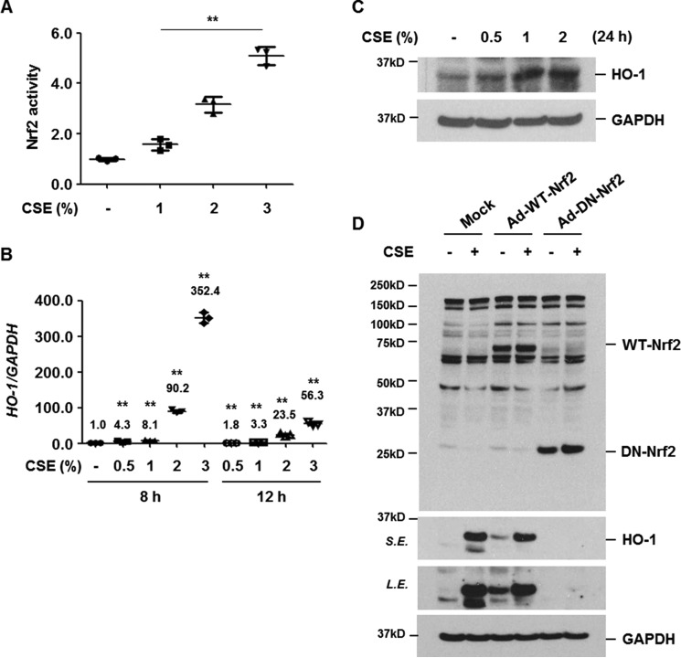
CSE increases the level of HO-1 expression via Nrf2 activation. A, BEAS-2B cells were treated with CSE (0, 1, 2, and 3%) for 4 h. Nrf2 activity using nuclear proteins was measured using a DNA-binding ELISA kit for activated Nrf2 transcription factor. Data represent the mean ± S.D. of triplicate experiments. B, BEAS-2B cells were stimulated with CSE (0, 0.5, 1, 2, and 3%) for 8 or 12 h. Total RNA was isolated and quantitative real-time PCR for HO-1 and GAPDH was performed. Data represent the mean ± S.D. of triplicates. **, p < 0.05. C, BEAS-2B cells were treated with CSE (0, 0.5, 1, and 2%) for 24 h. D, BEAS-2B cells were infected with control (Mock), wild-type Nrf2 (Ad-WT-Nrf2), or dominant-negative Nrf2 (Ad-DN-Nrf2) adenovirus vector. Forty-eight hours after infection, the cells were treated with CSE (1%) for 24 h. Total cellular extracts were subjected to Western blot analysis for HO-1, Nrf2, and GAPDH (C and D). The results are representative of three independent experiments. S.E., short exposure; L.E., long exposure.

