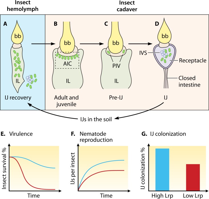
FIG 2.
Series of Steinernema host intestinal environments encountered by Xenorhabdus. (A to D) Simplified intestinal structures of nematodes (not whole organisms) at different stages (IJ, adult and juvenile, and pre-IJ) are schematically represented, with Xenorhabdus bacteria indicated by green ovals. (A) Once an IJ nematode enters the insect hemocoel from the soil, X. nematophila bacteria (green ovals) are released from the widening intestinal lumen (IL) of the recovering IJ into insect hemolymph during infection. (B) Adult and juvenile nematodes in the insect cadaver are colonized by symbiotic bacteria at the anterior intestinal cecum (AIC), a region within the intestine immediately below the pharyngeal intestinal valve (PIV). (C) In a pre-IJ nematode, a few symbiotic bacterial cells are enclosed in pouches within the PIV. (D) In an IJ nematode with a closed intestine, symbiotic bacteria colonize the receptacle. Bacteria either are associated with the intravesicular structure (IVS) or move freely in the receptacle lumen. (E) X. nematophila bacteria expressing low levels of Lrp are more virulent toward insects. Blue curve, high-Lrp-expressing bacteria; red curve, low-Lrp-expressing bacteria. (F) X. nematophila bacteria expressing high levels of Lrp better support nematode reproduction in the insect cadaver. Blue curve, high-Lrp-expressing bacteria; red curve, low-Lrp-expressing bacteria. (G) X. nematophila bacteria expressing high levels of Lrp show a high colonization frequency in IJ nematodes. bb, basal bulb; IL, intestinal lumen; AIC, anterior intestinal cecum; PIV, pharyngeal intestinal valve; IVS, intravesicular structure.

