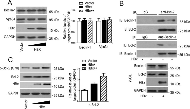
FIG 3.
HBx suppresses the association of beclin-1 with Bcl-2. (A) HepG2 cells were transfected with increasing doses of pHBx. At 48 h posttransfection, Western blot analysis was performed with the antibodies indicated. (Left panel) Levels of beclin-1 and VPS34 relative to the level of GAPDH were examined by densitometric analysis, and the value from empty-vector-transfected cells was set at 1.0 (n = 4). *, P < 0.05. (B) HepG2 cells were transfected with pHBx or the empty vector. At 48 h posttransfection, cell lysates were subjected to immunoprecipitation with anti-Bcl-2 or anti-beclin-1 antibody, followed by immunoblotting (IB) with anti-beclin-1 or anti-Bcl-2 antibody. (C) HepG2 cells were treated as described for panel A, Western blot analysis was performed with the antibodies indicated. (Left panel) Levels of p-Bcl2 relative to the level of GAPDH were examined by densitometric analysis, and the value from empty-vector-transfected cells was set at 1.0 (n = 4). *, P < 0.05. WCL, whole-cell lysate.

