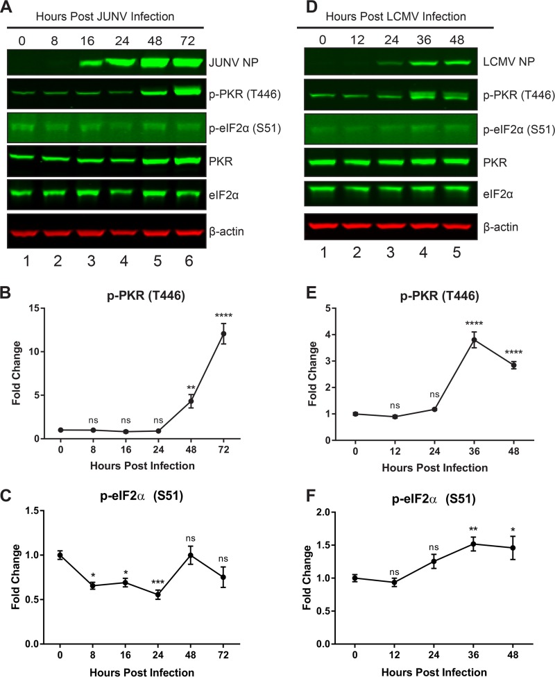
FIG 7.
PKR is activated following JUNV infection but cannot phosphorylate eIF2α. A549 cells were infected with JUNV (A to C) or LCMV (D to F). Infected cell lysates were collected during a time course of acute infection. (A and D) Viral NPs, phosphorylated PKR (T446), total PKR, phosphorylated eIF2α (S51), total eIF2α, and β-actin were visualized by Western blotting. Phosphorylated PKR (B and E) and phosphorylated eIF2α (C and F) were quantified and compared using one-way ANOVA. Data are presented as mean fold changes ± standard errors of the means (SEM) for 2 independent experiments featuring 3 technical replicates each. ns, not significant (P > 0.05); *, P ≤ 0.05; **, P ≤ 0.01; ***, P ≤ 0.001; ****, P ≤ 0.0001.

