FIG 6.
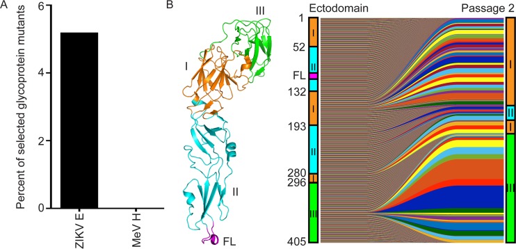
The ZIKV E gene is tolerant of insertional mutagenesis. (A) Comparison of ZIKV E and measles virus hemagglutinin (MeV H) insertional tolerances. The graph shows the percentage of insertional mutants in each glycoprotein coding sequence in the input library that were enriched >2 standard deviations above average following passaging (see Materials and Methods for analysis details). (B) (Left) Crystal structure (PDB entry 5IRE) of the E ectodomain monomer (48). The protein domains EDI (orange), EDII (cyan), EDIII (green), and fusion loop (FL) (magenta) are indicated. (Right) Viral progression plots showing the selection of insertion sites in passage 2. The linear representation of the E protein to the left of the plot is drawn to scale, while the illustration to the right is distorted to match the relative insertion abundance in each domain at passage 2.

