FIG 7.
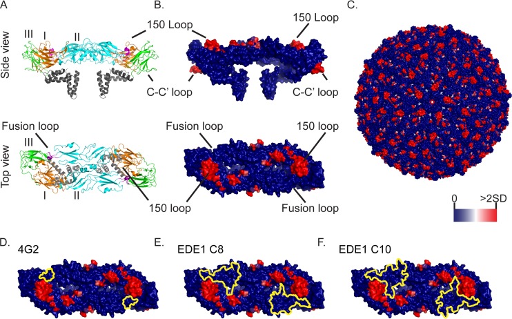
Mapping of insertion sites and broadly protective epitopes on the ZIKV E protein. (A) Top (as if viewed from outside the virion) and side views of the ribbon structure of the ZIKV E dimer complex crystal structure (PDB entry 5IRE) (48). The different protein domains are labeled and highlighted (EDI, orange; EDII, cyan; EDIII, green; fusion loop, magenta; and transmembrane domain and cytoplasmic tail, gray). The E protein 150-loop, fusion loop, and C-C′ loops are indicated. (B) The same crystal structure views, but with the transposon insertion frequencies at passage 2 indicated in colors from dark blue (0 reads with inserts) to red (2 standard deviations above average) on the surface structure of the E dimer complex. (C) Insertion frequencies mapped on the surface view of the whole ZIKV virion (48). (D to F) E protein structures showing both the passage 2 insertion frequencies (using the same heat map as that in panel C) and the binding sites (yellow outlines) of the 4G2 (D), C8 (E), and C10 (F) E antibodies. Antibody binding sites, which were previously mapped on the DENV E protein, were inferred by comparisons of the ZIKV and DENV protein sequences (27, 29).

