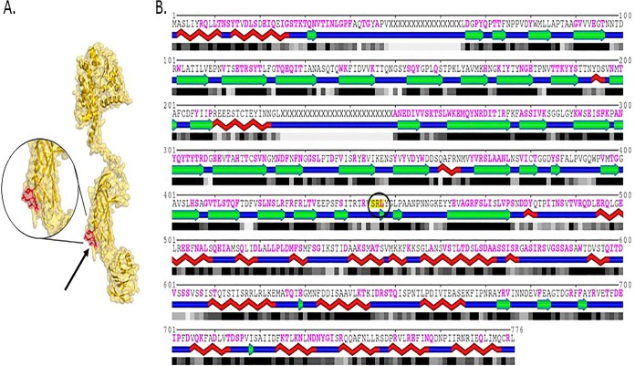
FIG 1.
RRV VP4 structure. (A) Cryo-electron microscopy (cryoEM)-resolved rotavirus particle structure (PDB code 4V7Q) showing the location of the SRL motif on the surface of the outer capsid protein VP4. The SRL-containing motif (residues 444 to 450; VSRLYGL) is shown in red on the surface of the isolated chain BX of structure 4V7Q (shown using yellow surface and backbone rendering). (B) One-dimensional profile of the same chain, with red braids, green arrows, and blue segments representing helices, beta strands, and loops, respectively, and shaded boxes representing the solvent accessibility of individual amino acid residues (with black corresponding to fully buried and gray to partially exposed residues). The location of the SRL motif (residues 445 to 447; highlighted in yellow and circled) within the BX chain of structure 4V7Q is indicated, with mapping of all protein interaction interfaces shown in magenta. As can be seen, the SRL motif is located in a generally accessible region. (Adapted from reference 33 with permission of the publisher.)

