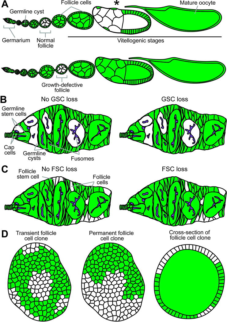
Fig. 2.

Diagrams of potential genetic mosaic analysis outcomes. (A) Top: Normal ovariole containing previtellogenic (“Normal follicle”) and vitellogenic (asterisk) follicles with GFP-negative germline cysts. Bottom: Follicle containing GFP-negative cyst shows a delay in growth, readily apparent in comparison to neighboring wild-type follicles. (B) A permanent clone derived from an identifiable GFP-negative GSC (left) populates the germarium (left). A recent GSC loss event is recognizable by the presence of GFP-negative germline cystoblasts/cysts within a mosaic germarium without the original GFP-negative mother GSC (right). (C) Permanent clones arising from an identifiable GFP-negative FSC in the germarium (left) or without a GFP-negative FSC (right), which indicates a loss event. (D) Transient (left) and permanent (middle) follicle cell clones are imaged in single planes for quantification of follicle cell proliferation by clone size or EdU incorporation frequency, respectively. Cross-sections of follicle cell clones (right) in the ovariole are often visible during germline cyst analyses.
