Fig. 2.
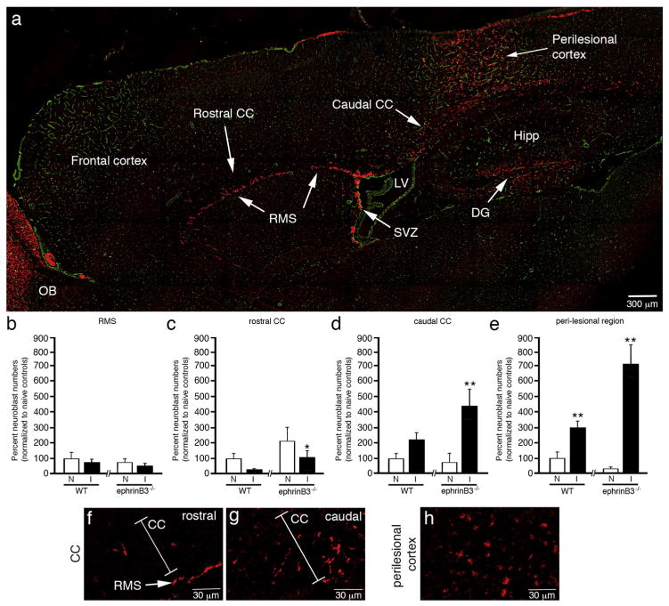
EphrinB3 increases neuroblast migration outside the SVZ and RMS. Immunolabeled sagittal brain section from a WT mouse showing anti-DCX labeled neuroblasts (red) in RMS, CC and perilesional cortex 3 days after CCI injury, while anti-CD31 (green) labeled vessels were used for tissue referencing (a). Stereological cell counts of the RMS (b), rostral CC (c), caudal CC (d) and peri-lesional cortex (e) show a rostral-to-caudal gradient of neuroblasts from the rostral CC towards the caudal CC and injury site. Increased neuroblasts numbers were observed in the rostral CC (c), caudal CC (d) and perilesional cortex (e) in ephrinB3−/− as compared to WT mice. High-magnification images of neuroblasts in the rostral CC (f), caudal CC (g) and perilesional cortex (h). CC, corpus callosum; DG, dentate gyrus; Hipp, Hippocampus; LV, lateral ventricle; OB, olfactory bulb; RMS, rostral migratory stream; SVZ, subventricular zone. *p < 0.05, **p < 0.01 as compared with their respective non-injured controls.

