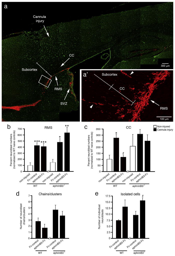
Fig. 3.
EphrinB3-Fc infusion reduces neuroblast migration into the overlying CC at 2 days following cannula injury. Immunolabeled sagittal brain section from a WT mouse shows anti-DCX labeled neuroblasts (red) in the RMS, CC and subcortical tissues at 2 days post-injury, while anti-CD31 (green) labeled vessels were used for tissue referencing (a). High-magnification inset of neuroblasts migrating from RMS to CC and subcortical tissues (arrowheads depict neuroblast chains) (a′). Stereology shows increased numbers of neuroblasts in the RMS (b) and CC (c) in Fc-control WT mice as compared with non-injured WT mice. Infusion of ephrinB3-Fc reduced neuroblast numbers in the CC of WT mice (c). EphrinB3−/− mice show increased chain (d) and isolated cell (e) migration in the rostral CC as compared with WT mice. Infusion of clustered ephrinB3-Fc reduced the number of neuroblast chains/clusters but increased the number of isolated neuroblasts. CC, corpus callosum; RMS, rostral migratory stream; SVZ, subventricular zone. *p < 0.05, **p < 0.01, ***p < 0.001 as compared with their respective non-injured controls; #p < 0.05 as compared with their respective Fc controls.

