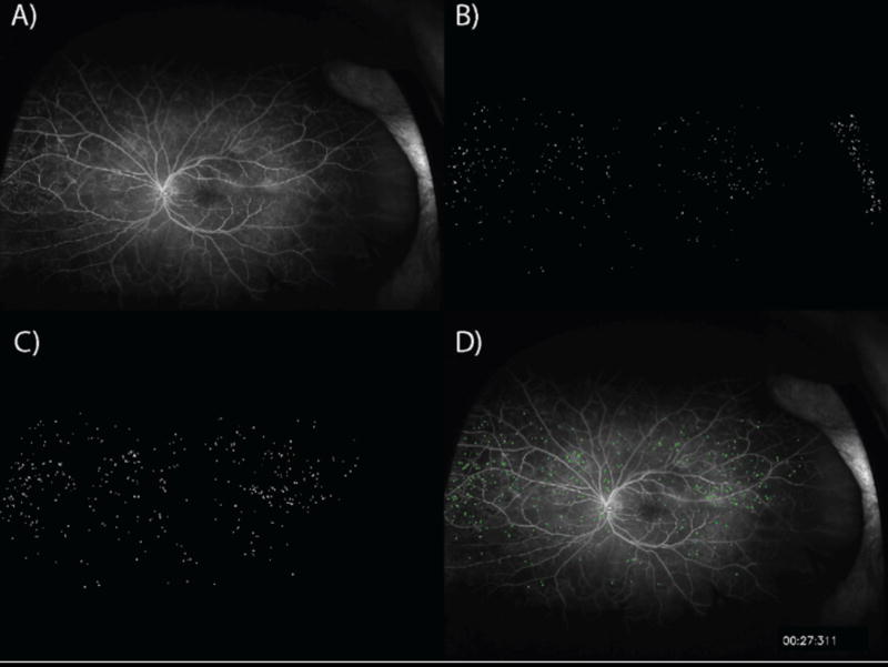
Figure 1. Automated microaneurysm detection.
A) An early time point ultra-widefield fluorescein angiogram image is selected. B) Initial detection of candidate microaneurysms (MAs). C) Candidate MAs are filtered based on peripheral intensity gradient. D) MAs are superimposed upon original early phase image.

