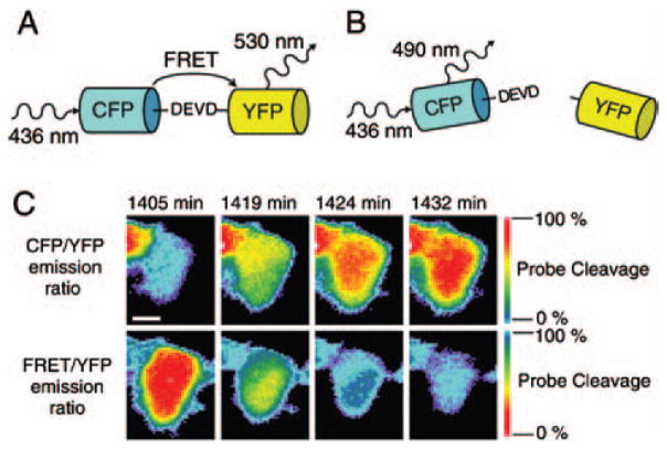
Fig. 5.
DEVD FRET probe containing DEVD as the specific cleavage site for caspase-3, Cyan Fluorescent Protein (CFP) as the FRET donor, and Yellow Fluorescent Protein (YFP) as the FRET acceptor. (A) Without the presence of caspase-3, CFP and YFP are linked by DEVD peptide. Upon CFP excitation, the energy was transferred to YFP by FRET. (B) With the presence of caspase-3, the DEVD linker was cleaved and the FRET between CFP and YFP was disappeared. (C) DLD-1 cell expressing DEVD FRET probe was treated with 1 μM staurosporine for inducing apoptosis. The changes of CFP/YFP and FRET/YFP emission ratio indicate the cleavage of the DEVD FRET probe. Reproduced with permission from [129], C. L. O’Connor et al., Intracellular signaling dynamics during apoptosis execution in the presence or absence of X-linked-inhibitor-of-apoptosis-protein. Biochim. Biophys. Acta. 1783, 1903 (2008). © 2008.

