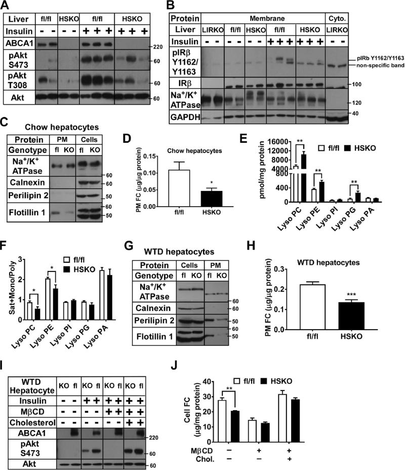
Figure 5. Primary hepatocyte plasma membrane (PM) free cholesterol (FC) content modulates insulin signaling. See also Figure S5.
Mice consuming a WTD for 16–24 weeks received portal vein saline (n=2 per genotype) or insulin (0.5 U/kg body weight; n=3 per genotype) injection; 5 min later, livers were harvested. (A) Liver Western blots. (B) Western blots of hepatic membrane-associated and cytosolic proteins. LIRKO, liver-specific insulin receptor knockout. (C–D) Primary hepatocytes from chow-fed mice (n=2 per genotype) were used to purify PM fractions (n=3 per treatment) for Western blot (C) and FC measures (D). (E–F) Liver lipids were extracted from chow-fed mice (n=4 per genotype) for lipidomic analyses. (G–H) Primary hepatocytes from WTD-fed mice (n=2 per genotype) were used to purify PM fractions (n=6 per treatment). Western blotting and lipid analyses: as in panels C–D. (I) Primary hepatocytes from WTD-fed mice (n=2 per genotype) were FC depleted (+MβCD) and repleted (+cholesterol) before insulin stimulation (n=3 per treatment) and Western blotting. (J) Cells from panel I were lipid extracted and FC measured. Results are representative of 2–3 separate experiments.

