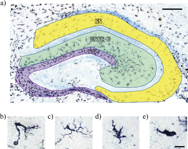
Figure 1. Hippocampal subdivisions and microglia morphologies.

Hippocampal subdivisions and representative images of microglial morphological categories used in cell counting. The hippocampus was divided into the dentate gyrus (DG; purple), the molecular layer/stratum radiatum (SR/ML; green), and the stratum oriens (SO; yellow). Microglia morphology was assessed by classifying microglia as phagocytic (b), ramified (c), transitioning (d), or amoeboid (e) following immunohistochemical staining for the pan-microglial marker, Iba1. Scale bar = 200 μm for panel (a), and 12.5 μm for panels (b)–(e).
