
Figure 3. Differences in microglial number across hippocampal sub-region and sex differences within sub-regions.
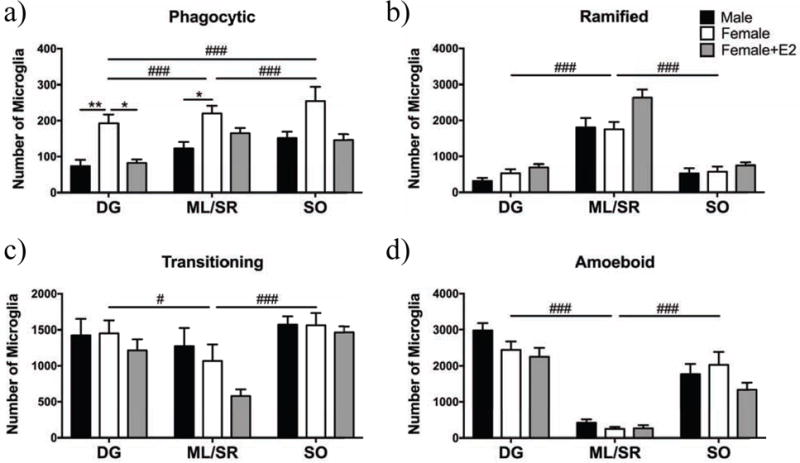
Microglia morphology was classified as phagocytic and/or transitioning, ramified or amoeboid across three sub-regions of the hippocampus in males (black), females (white), and females treated with E2 (gray). There were more phagocytic microglia in the ML/SR and SO compared to the DG, and more phagocytic microglia in the SO compared to the ML/SR (a). Females had significantly more phagocytic microglia compared to males in the DG and ML/SR (a). Females had more phagocytic microglia in the DG compared to estradiol-treated females (a). There were more ramified microglia in the ML/SR compared to the SO and DG (b). There were more transitioning microglia in the DG and SO compared to the ML/SR (c). There were more amoeboid microglia in the DG and SO compared to the ML/SR (d). DG = dentate gyrus, ML/SR = molecular layer/stratum radiatum, and SO = statrum oriens. * p < 0.05, ** p < 0.01 for pairwise comparisons within areas/morphologies; # p < 0.01, ### p < 0.001 for comparisons between areas independent of sex.