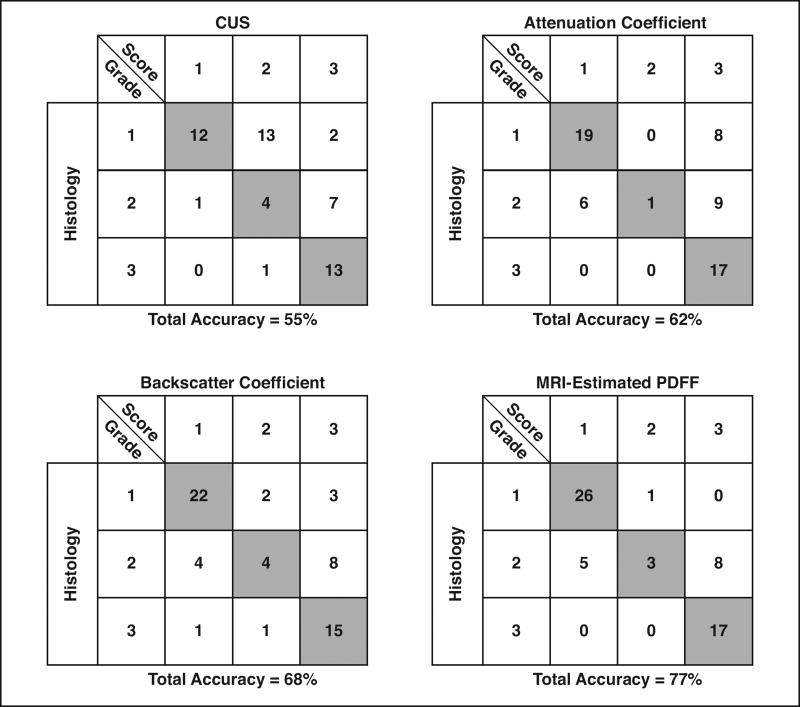
Fig. 3.
Classification tables showing number of correctly classified (shaded boxes) and incorrectly classified (unshaded boxes) patients for each histologic steatosis grade for each imaging measure. Tables represent Classification data based on two-radiologist consensus conventional ultrasound (CUS) scores, two-analyst mean quantitative ultrasound scores (attenuation and backscatter coefficients), or MRI-estimated proton density fat fraction (PDFF) scores.

