
Fig. 4.
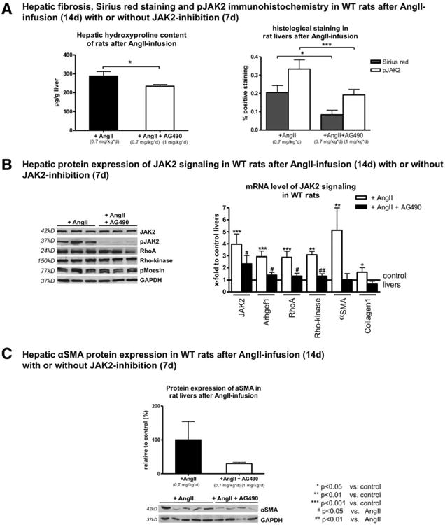
Stimulation of AT1R with AngII activated JAK2, but JAK2 inhibition attenuated hepatic fibrosis. (A) Hepatic hydroxyproline content and quantification of Sirius red and pJAK2 IHC staining are shown in rats after AngII infusion (0.7 mg/kg/day) for 14 days with or without additional AG490 (1 mg/kg/day) 7 days before sacrifice. JAK2 inhibition with AG490 blunted fibrosis development in these rats (for representative sections, see Supporting Fig. S7). (B) Protein levels of AT1R, JAK2, pJAK2, Arhgef1, RhoA, Rho-kinase, and pMoesin were analyzed using western blotting, and mRNA levels of JAK2, Arhgef1, RhoA, Rho-kinase, α-SMA, and Col1 were analyzed by RT-PCR in liver samples of rats after AngII infusion (0.7 mg/kg/day) for 14 days with or without additional AG490 (1 mg/kg/day) 7 days before sacrifice. (C) Hepatic protein levels of α-SMA are shown in rats after AngII infusion (0.7 mg/kg/day) for 14 days with or without additional AG490 (1 mg/kg/day) 7 days before sacrifice.