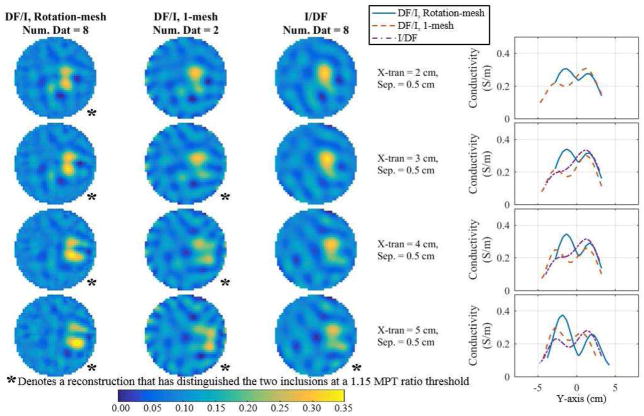
Fig. 8.
Reconstructions comparing DF/I to the I/DF technique for 8 fused datasets corresponding to the DF/I’s smallest distinguishable separation at each x-translation. The first three columns show the reconstructions and the final column shows the corresponding profiles through the peaks. If there is no profile for a number of fused datasets then two peaks were not detected.

