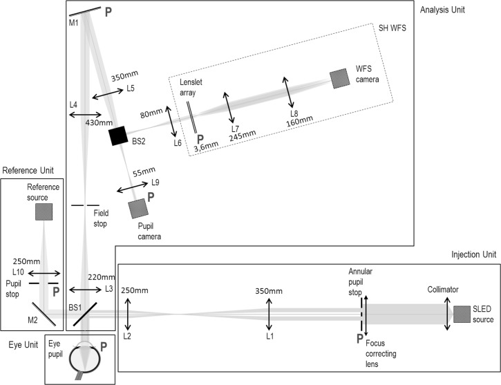
Fig. 1.
Schematic drawing of the experimental set-up, comprising a Reference Unit (used to acquire the reference wavefront), an Injection Unit creating a point source on the retina and an Analysis Unit with a custom-made Shack-Hartmann Wavefront Sensor (SH WFS), in parallel with a pupil camera.(L: lens - associated focal lengths are reported on the schematic drawing, BS: beam splitter, M: mirror). All pupil planes (marked with P) are optically conjugated.

