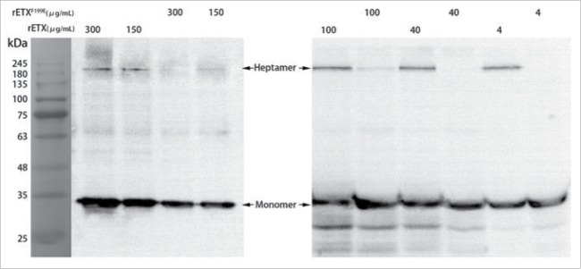
Figure 5.
Analysis of heptamer formation by rETX and rETXF199E. MDCK cells were treated with 200 μl of toxins (4 μg/mL to 300 μg/mL) for 30 min at 37°C. Samples were solubilized, and analyzed by SDS-PAGE, and then immunoblotted with an anti-His monoclonal antibody and a HRP-coupled goat anti-mouse IgG antibody (1:50,000). The result was photographed using an AE-1000 cool CCD image analyzer.

