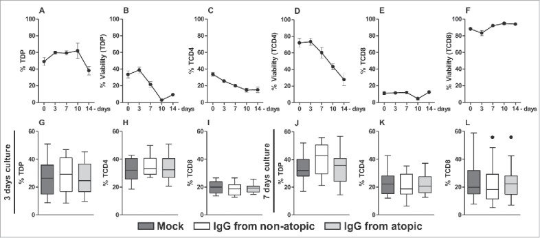
Figure 1.

Time course of cell frequency and viability and of the effect of purified IgG on human intra-thymic αβT cells. Thymocytes from children less than 7 d old (n = 14) were evaluated at time 0 or after 3, 7, 10 and 14 d in culture in RPMI medium supplemented with FBS. At each time point, the frequency and viability of TDP (A-B), TCD4 (C-D) and TCD8 cells (E-F) were evaluated by flow cytometry. Thymocytes were also cultured for 3 or 7 d in RPMI medium supplemented with FBS in the absence (mock) or presence of 100 µg/mL IgG purified from atopic or non-atopic individuals. At each time point, the frequency and viability of TDP (G and J), TCD4 (H and K) and TCD8 cells (I and L) were evaluated by flow cytometry. The symbols represent the means with standard error. The results are illustrated by box and whiskers graphs with 25th percentiles, and the Tukey method was used to plot outliers.
