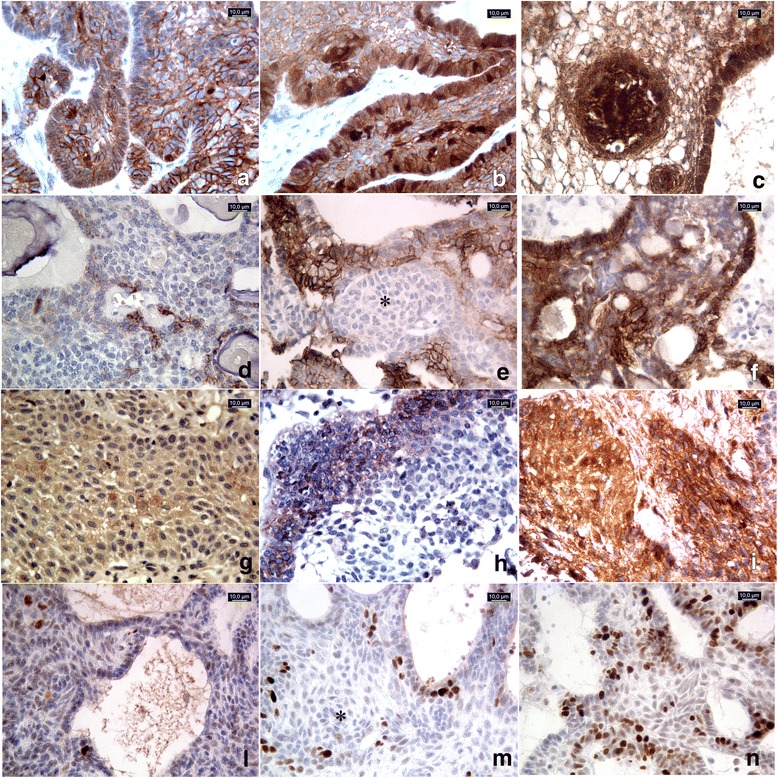
Fig. 1.

45 cases were examined with β-catenin, Ki67 and pATM antibodies, 30 cases with CD133 and 31 cases with CD166. (a-c) β-catenin immunostaining score: moderateand strong nuclear signal in <10% (a, adaCPnr23of Table 1) and in 10–50% (b, adaCPnr40) of neoplastic cells, respectively. The signal was more represented in cells forming “whirl-like” structures (c, adaCPnr7). d-f CD166immunostaining score: strong membranous signal observed in <10% of neoplastic cells (d, adaCPnr8); moderate signal observed in 10–50% of cells (e, adaCPnr27), sparing the “whirl-like” structures (*). Strong reactivity in >50% of neoplastic cells (f, adaCPnr31). g-i CD133immunostaining score: signal with moderate intensity observed in 10–50% of neoplastic cells in papCP (g, papCPnr14) and in adaCP (h, adaCPnr44); strong and diffuse (51–80% of neoplastic cells) immunoreactivity in papCP (i, papCPnr13). L-n Ki67 Labeling index: low proliferative index Ki67 (<5%) (l, adaCPnr3); high Ki67 L.I sparing cells forming “whirl-like” clusters (*) (m, adaCPnr15); Ki67 L.I. calculated as 25% (n, adaCPnr7). All pictures were captured at 40× magnification
