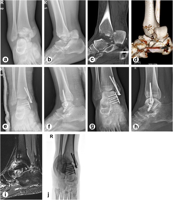
Fig. 3.

Examples of images obtained from case 18 (A 23-year-old female patient with a right ankle injury due to heavy object crushes). a–b Anterioposterior and lateral view of X-ray before operation. c–d CT and 3D reconstruction before operation. e–f X-ray at 7 days after operation. g–h Anterioposterior and lateral view of X-ray at 6 months follow-up. Subtalar arthritis was obviously seen. i MRI at 1 year follow-up. Talocrural and subtalar arthritis were obviously seen. j Canale view of X ray at 1 year follow-up
