Fig. 9.

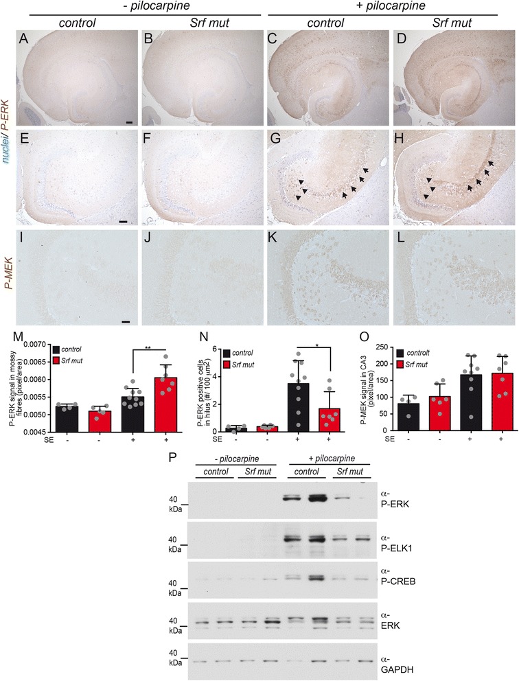
Activity of MAP kinase signaling members is altered upon SRF ablation. a-h Hippocampal sections were labeled with anti P-ERK directed antibodies and counterstained with Nissl. Without SE induction (−pilocarpine), almost no P-ERK signals were observed in heterozygous (a, e) or Srf mutant (b, f) animals. In heterozygous animals with SE, P-ERK levels in the hilus were upregulated (arrowheads in g) in contrast to Srf mutant animals (arrowheads in h). In addition, P-ERK levels were augmented in the CA3 region of epileptic control animals (c; arrows in g). This was even further increased upon SRF ablation and now P-ERK co-localized with mossy fibers (arrows in h). e-h are higher magnifications of (a-d). i-l P-MEK levels were low in heterozygous (i) and SRF deficient (j) animals without SE. Pilocarpine injection induced P-MEK in a similar manner in heterozygous (k) and mutant (l) animals. m-o Quantification of P-ERK levels in mossy fibers (m) and hilus (n) as well as of P-MEK (o). Data are represented as mean ± SD. Individual animals are labeled with grey circles. p Immunoblotting of heterozygous and SRF-deficient hippocampal lysates derived from animals without (−pilocarpine) or with 40 min SE (+pilocarpine). For each condition, two animals are depicted. P-ELK1, P-ERK and P-CREB levels were elevated by an SE in heterozygous control animals but not as much in Srf mutant animals. Scale-bar a-d = 200 μm; e-h = 75 μm; i-l = 100 μm
