FIGURE 4.
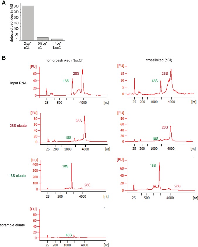
Specific RNP captures of 18S and 28S rRNAs from HeLa cells. (A) HeLa cells were UV-irradiated and poly(A) RNA was subsequently purified with oligo(dT). Two micrograms or 0.5 µg of eluted RNA was analyzed by mass spectrometry. A nonirradiated sample (NocCl) was used as control. The number of identified peptides is shown. (B) RNA eluates using probes against 28S rRNA or 18S rRNA were analyzed by bioanalyzer. Panels show the electropherograms of input (upper panels), 28S capture (middle upper panels), 18S capture (middle bottom panels), and capture with LNAscr of one representative experiment. The two peaks observed in the input samples correspond to 18S (2000 nt) and 28S (4000 nt) rRNAs. The y-axes display the fluorescence intensity derived from the dye labeling the RNA.

