Figure 1.
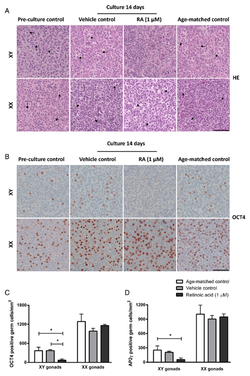
Morphology and presence of germ cells in human fetal gonad tissue cultured in hanging drop cultures. Human fetal testis and ovary tissue aged between gestational week (GW) 7-9 cultured for 2 weeks with or without addition of retinoic acid (RA) in the media and compared to pre-culture and age-matched controls (not cultured). The age-matched control corresponds to the age of fetal samples at the time of experimental start plus two weeks. Tissue from 4-7 fetuses of each gender was investigated. A: Haematoxylin and eosin (HE) staining. Arrows point to gonocytes (in XY panel) and oogonia (in XX panel), scale bar corresponds to 50 µm. B: Immunohistochemical staining with the pluripotency marker octamer-binding transcription factor 4 (OCT4) that stains gonocytes in human fetal testis and oogonia/oocytes in human fetal ovaries. Counterstaining with Mayer haematoxylin, scale bar corresponds to 50 µm. C: Quantification of germ cells determined as the number of germ cells stained with OCT4 per mm2. Values represent mean ± sd. * indicates significant difference (p < 0.05). D: Quantification of germ cells determined as the number of germ cells stained with transcription factor AP-2 gamma (AP2γ) per mm2. Values represent mean ± sd. * indicates significant difference (p < 0.05). Abbreviations: RA, retinoic acid; XX, fetal ovaries; XY, fetal testes.

