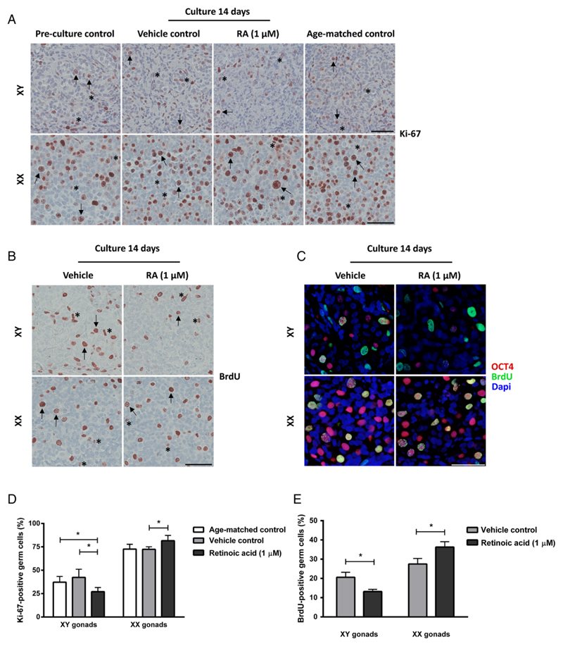
Figure 2.
Proliferation of germ cells and somatic cells in human fetal gonad cultures. Fetal testis and ovary tissue aged between gestational week (GW) 7-9 cultured for 2 weeks with or without presence of retinoic acid (RA) in the media and compared to pre-culture and age-matched controls (not cultured). The age-matched control corresponds to the age of fetal samples at the time of experimental start plus two weeks. Tissue from 4-7 fetuses of each gender was investigated. A: Immunohistochemical staining with the proliferation marker Ki-67 antigen (Ki-67). Arrows point to proliferating (Ki-67 positive) gonocytes (in XY panel) and oogonia (in XX panel), asterisk marks proliferating somatic cells. Counterstaining with Mayer haematoxylin, scale bar corresponds to 50 µm. B: Immunohistochemical staining with the proliferation marker 5’-bromo-2’-deoxyuridine (BrdU). Arrows point to proliferating (BrdU positive) gonocytes (in XY panel) and oogonia (in XX panel), asterisk marks proliferating somatic cells. Counterstaining with Mayer haematoxylin, scale bar corresponds to 50 µm. C: Immunofluorescenct staining with octamer-binding transcription factor 4 (OCT4) (red) and BrdU (green). Counterstaining with 4',6-diamidino-2-phenylindole (DAPI) (blue), scale bar corresponds to 50 µm. D: Quantification of proliferating germ cells determined as the percentage of germ cells stained with Ki-67. Values represent mean ± sd. * indicates significant difference (p < 0.05). E: Quantification of proliferating germ cells determined as the percentage of germ cells stained with BrdU, based on immunohistochemical stainings. Values represent mean ± sd. * indicates significant difference (p < 0.05). Abbreviations: RA, retinoic acid; XX, fetal ovaries; XY, fetal testes.

