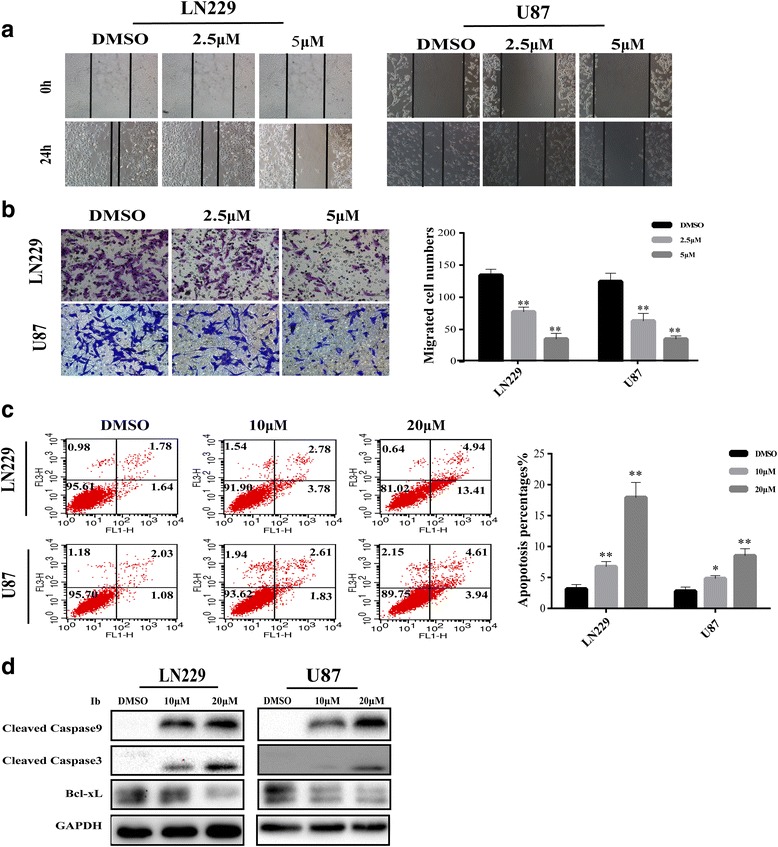
Fig. 2.

Ibrutinib suppresses cell migration and induces apoptosis in GBM cells. (a) The migratory ability of LN229 and U87 cells was evaluated in a wound healing assay with cells treated with various concentrations of ibrutinib for 24 h. (b) The results of trans-well assay with LN229 and U87 cells treated with different concentrations of ibrutinib for 24 h. Statistical analyses of the migrated cells are shown on the right; **p < 0.01. (c) The percentage of apoptotic cells in LN229 and U87 cell population treated with increasing concentrations of ibrutinib, as detected by flow cytometry with annexin V-PI staining. Data are shown as the mean ± SD and are from three independent experiments; *p < 0.05, **p < 0.01. (d) The expression of apoptosis-associated proteins cleaved caspase 9, cleaved caspase 3, and Bcl-xL were detected by western blotting following the treatment of cells with increasing concentrations of ibrutinib for 48 h
