
Fig. 3.
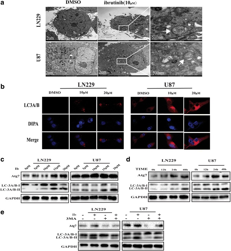
Ibrutinib induces autophagy in GBM cells. (a) TEM revealed autophagosome ultrastructures in the enlarged images (arrows) after a 24-h treatment with 10 μM ibrutinib. (b) Representative images of immunocytochemistry. Red fluorescence indicates the presence of LC-3 protein. (c, d) GBM cells were incubated with different concentrations of ibrutinib for 24 h (c) or with 10 μM ibrutinib for various times (d), and LC3A/B-II, Atg7, and GAPDH levels were assessed by immunoblotting. (e) LC3A/B and Atg7 levels examined by western blot analysis in LN229 and U87 cells after treatment with ibrutinib (10 μM) or DMSO, in the absence or presence of 3MA (2 nM)