
Fig. 6.
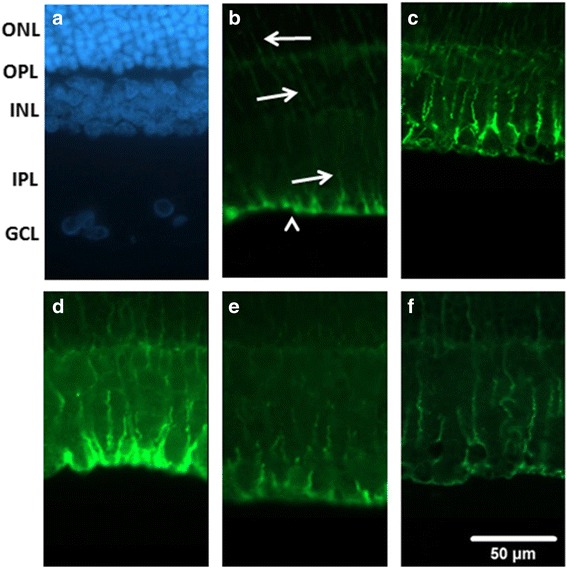
Vimentin (green) immunohistochemistry. a shows the cellular nucleus of the sham retina labeled with 4,6-diamidine-2-phenylindole dihydrochloride (DAPI, blue). b shows a sham retina with the vimentin immunoreactivity at the end feet (arrow heads) in the GCL and at the processes in the IPL, INL and ONL (arrows). c shows a retina that received I/R and preischemia adminstration of vehicle; in contrast to the sham retina, the anti-vimentin immuolabeling has increased. (d, e, f) are sections from retinas that received I/R and preischemia administration of 1.35 g/kg/day XFZYD (d, XFZYD1.35 + I/R), 2.7 g/kg/day XFZYD (e, XFZYD2.7 + I/R) or postischemia administration of 2.7 g/kg/day XFZYD (f, I/R + XFZYD2.7). Moreover, this ischemia-induced alteration is clearly and dose-dependently reduced when the ischemic retina is preadministrated with 1.35 and 2.7 g/Kg/day XFZYD. Postischemia administration of 2.7 g/Kg/day XFZYD also clearly attenuated this ischemia-induced alteration. ONL: outer nuclear layer, OPL: outer plexiform layer, INL: inner nuclear layer, IPL: inner plexiform layer, GCL: ganglion cell layer. Scale bar = 50 μm