Fig. 8.

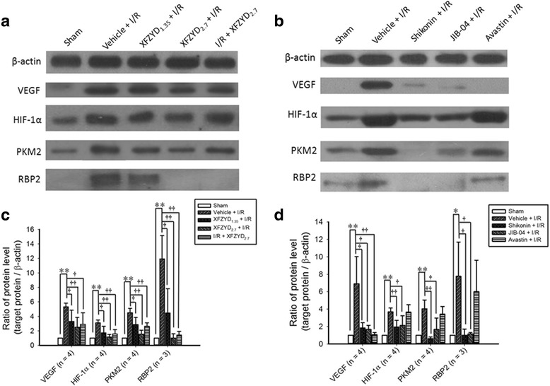
The ratios of the protein concentrations of VEGF, HIF-1α, PKM2 and RBP2 relative to β-Actin. a and b showed the Western blotting for expressions of β-actin, VEGF, HIF-1α, PKM2 and RBP2. In figure a, Lane 1 is a retina that received the sham procedure (Sham); Lane 2 is the vehicle-preadministered ischemic retina (Vehicle + I/R); Lanes 3 ~ 5 are from retinas that received ischemia plus reperfusion and were preadministrated with 1.35 g/kg/day XFZYD (XFZYD1.35 + I/R), 2.7 g/kg/day XFZYD (XFZYD2.7 + I/R) or postadministrated with 2.7 g/kg/day XFZYD (I/R + XFZYD2.7). In figure b, Lane 1 is from a sham retina (Control); Lane 2 is the vehicle-preadministered ischemic retina (Vehicle + I/R); Lanes 3 ~ 5 are from retinas that received ischemia plus reperfusion and were preadministrated with 4 μM Shikonin (PKM2 inhibitor), 10 μM JIB-04 (RBP2 inhibitor) and 100 mg/4 ml Avastin (anti-VEGF). (c and d) showed the protein levels of VEGF (n = 4), HIF-1α (n = 4), PKM2 (n = 4) and RBP2 (n = 3) divided by the level of β-actin. */** or †/†† indicates significance (p < 0.05/p < 0.01; pictures (c and d): Sham vs. Vehicle + I/R) or significance (p < 0.05/p < 0.01; picture c: Vehicle + I/R vs. XFZYD1.35 + I/R, XFZYD2.7 + I/R, or I/R + XFZYD2.7; Picture d: Vehicle + I/R vs. Shikonin + I/R, JIB-04 + I/R, or Avastin + I/R). Each bar indicates the mean values ± SD. VEGF: vascular endothelial growth factor, HIF-1α: hypoxia inducible factor 1α, PKM2: pyruvate kinase M2, RBP2: retinoblastoma-binding protein 2
