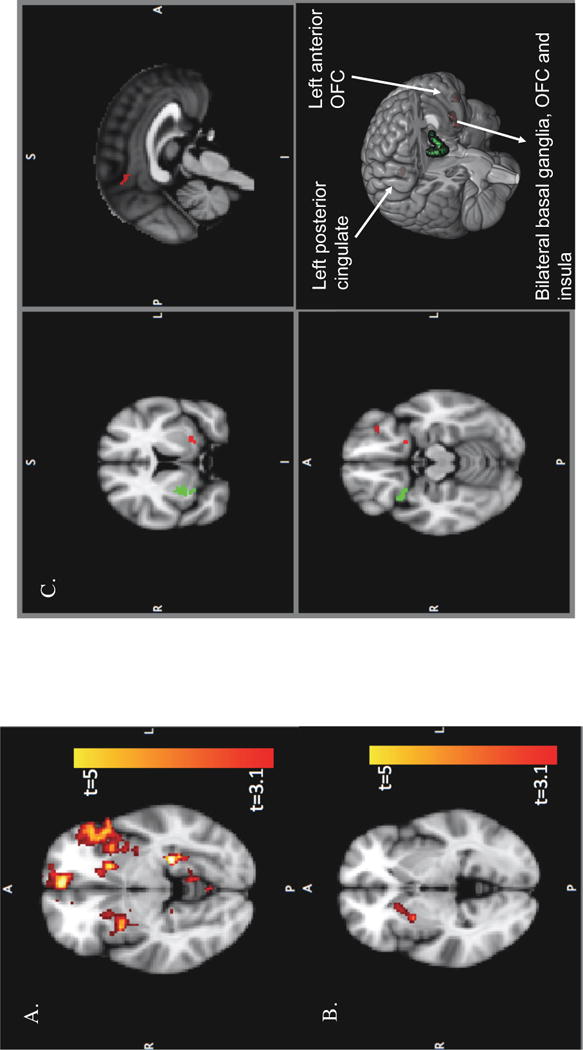
Figure 3. Neural Circuitry of Mental Representations of the Deceased.

A. Clusters showing greater response to deceased-related as compared to control stories (D_STO>CLD_STO). Clusters are significant at a voxel-wise threshold of t523=3.1, p<0.001 and cluster corrected with a threshold of p<0.05. A large cluster in right insula as well as a distributed network across anterior and posterior cingulate, orbital prefrontal cortex and bilateral basal ganglia are seen. Image coordinates (X,Y,Z=25,70,33) B. Cluster in right putamen, insula, and caudate whose activity is associated with viewing pictures of the deceased as compared to controls (D_PIC>CLD_PIC) of t522=3.1, p<0.001, cluster-p <0.05. Image coordinates (X,Y,Z=60,67,35) C. Red and green maps together display clusters in bilateral basal ganglia, OFC and insula as well as left anterior OFC and posterior cingulate conjointly activated by pictures and stories of the deceased as compared to the control conditions ((D_STO>CLD_STO) AND (D_PIC>CLD_PIC)) (voxel p<0.001, cluster-p <0.1). Green mask highlighted by 3D rendering displays the larger cluster on the right that survived a more stringent cluster size threshold of p<0.05 and was previously seen in cluster corrected story and picture results. Image coordinates (X,Y,Z=45,67,28)
