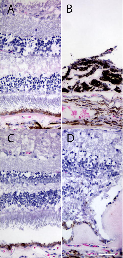
Figure 3.
Histology of the right (A,B) and left (C,D) eyes from subject 13. In areas of the retina remote from the deposits, the neural retina and RPE were relatively normal. Some subretinal material was observed regionally (e.g., C). The OD possessed an atypical neovascular membrane (lasered previously) that was heavily infiltrated with pigmented cells (B); the neural retina above this deposit was detached and atrophic. Where large deposits were present (D), the retina showed remodeling that included photoreceptor tubulations and loss of the outer nuclear layer. Scalebar = 50µm.

