
Fig. 1.
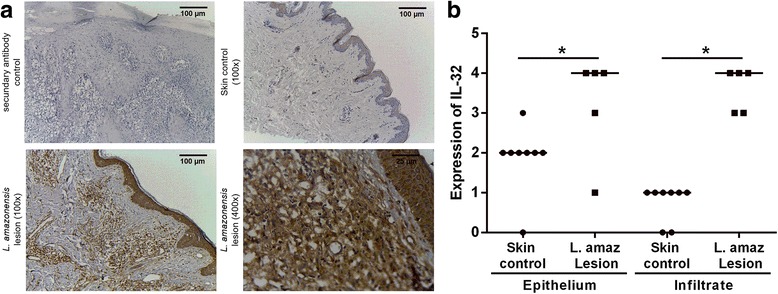
Expression of IL-32 in American tegumentary leishmaniasis lesions caused by L. amazonensis. a Fragments of lesions from ATL patients infected with L. amazonensis and skin from healthy controls were included in paraffin and submitted to immunohistochemistry for IL-32. The reaction was revealed with 3,3′-Diaminobenzidine and Meyer’s hematoxylin used to counterstain. b Evaluation of IL-32 expression score was determined according to the percentage of cells expressing IL-32. The scores represent: 0 (absence of stained cells), 1 (1–25% of stained cells), 2 (26–50% stained cells), 3 (51–75% stained cells), and 4 (76–100% stained cells). *P < 0.05 (Mann-Whitney test)