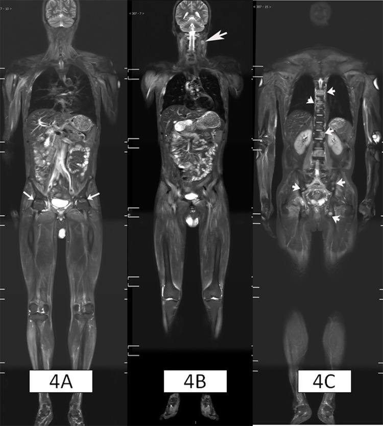
Fig 4. 43-year-old male patient, displaying DM with nasopharyngeal cancer and cervical lymph nodes metastases, multiple bone metastases and bilateral femoral head necrosis.
Figure 4A showed the avascular necrosis at bilateral femoral head (white arrows); and Figure 4B showed swollen lymph node on the left side of the neck (white arrow); Figure 4C showed patchy abnormal high signals in the thoracic and lumbar spine and pelvis (white arrow). Vertebral biopsy confirmed skeletal metastases.

