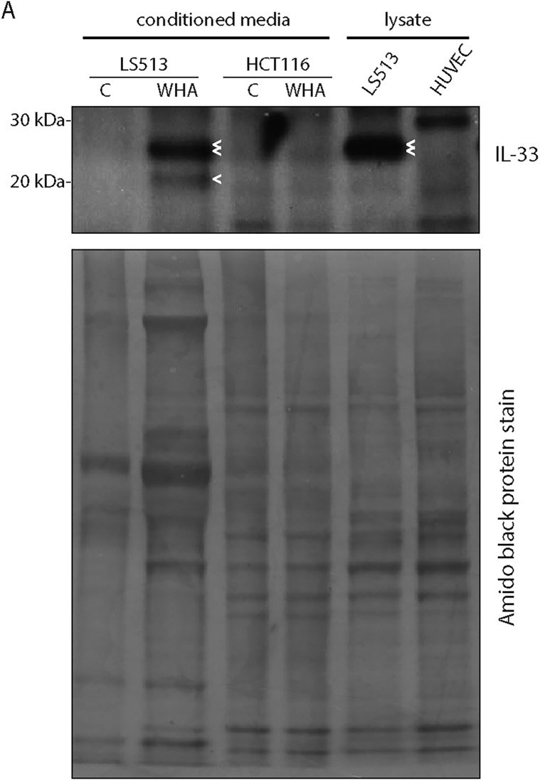
Fig 9. LS513 or HCT116 cells were cultured to confluence, changed into serum-free media and confluent cells “wounded” with pipette tip (wound healing assay, WHA) or not (control, C).
After 24h, conditioned media was collected for TCA precipitation of released proteins. Alternatively, LS513 or HUVEC cells were cultured to confluence, and lysates prepared using standard procedures, as a positive control. Media precipitate and lysates were analysed by Western blotting for expression of released or endogenous IL-33 isoforms. Subsequently, the blot membrane was stained with amido black to show protein loading in each lane. Double arrow denotes LTR-IL-33, single arrow denotes cleavage product.

