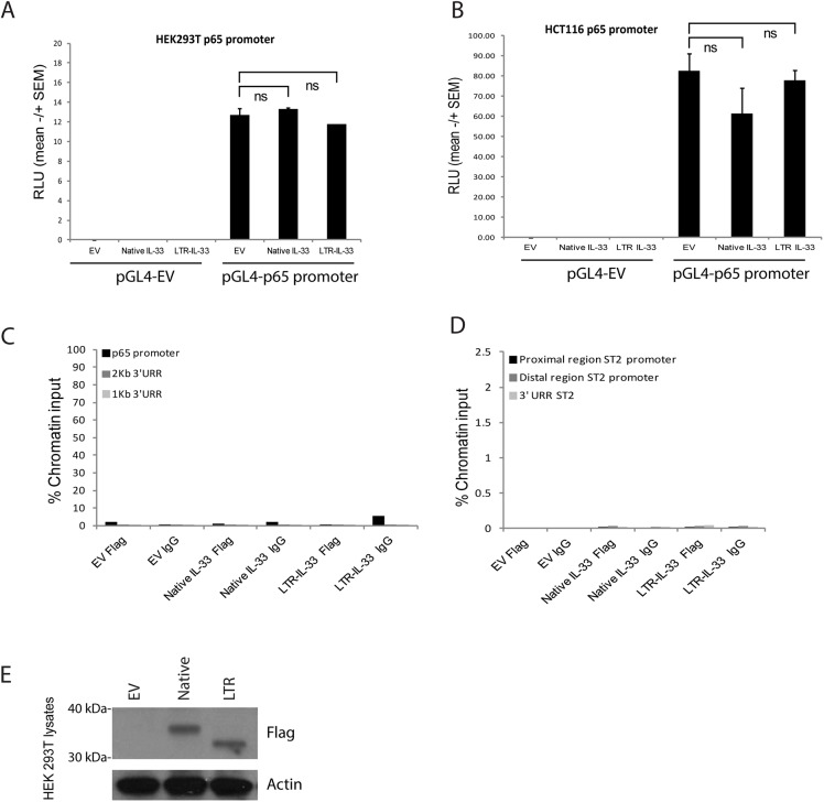
Fig 10.
A: HEK 293T or (B) HCT116 cells were transfected to express exogenous Native or LTR-IL-33 (or Empty Flag Vector control) and the full length p65 promoter region in the luciferase construct pGL4 (or the pGL4 Empty Vector control) and Renilla transcription control. After 43h, Relative Luciferase Units were assessed as a measure of p65 promoter activity. C, D: 293T HEK were transfected to express exogenous Native or LTR-IL-33 (or Empty Flag Vector control). 48h later, Chromatin immunoprecipitation was carried out using anti-FLAG antibodies or normal mouse IgG. Genomic DNA was amplified by PCR using primers (C) spanning the p65 promoter region. For the negative control region, genomic DNA was amplified using primers specific to two upstream UnRelated Regions (1kB or 2kB). D: specific to regions (proximal or distal) of the ST2 promoter, or a downstream UnRelated Region. E: HEK293T lysates from cells used in C and D were assessed by Western blotting using antibodies raised against Flag or Actin (loading control). Representative data of 3 independent experiments is shown.

