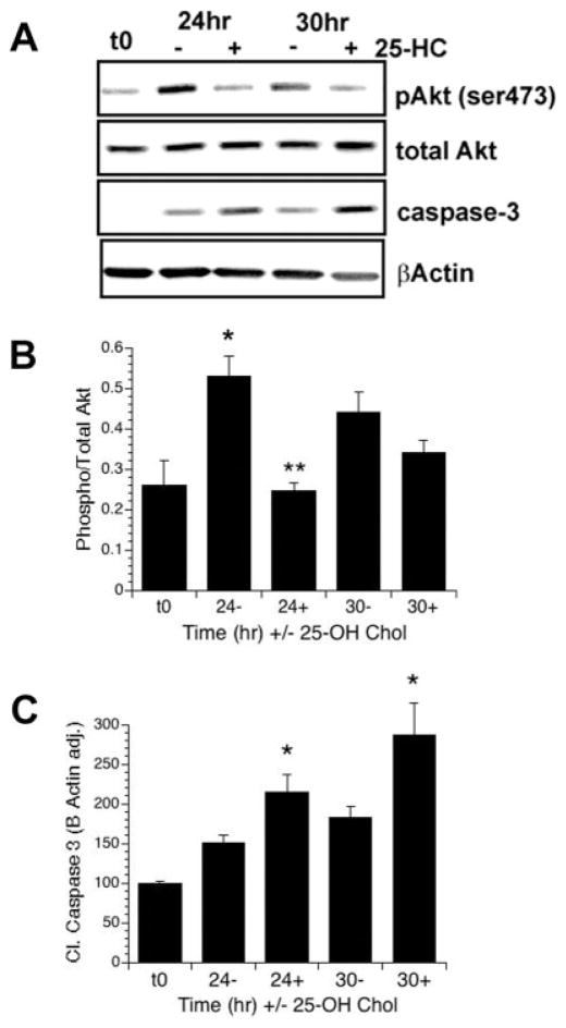
Fig. 3.
Inhibition of cholesterol biosynthesis blocks IGF-I-mediated sustained Akt phosphorylation. A–C: OPCs were treated with 10 ng/ml IGF-I in the presence (+) or absence (−) of 25-hydroxycholesterol (25-HC) for 24 or 30 hr in serum-free media. A: Representative Western blots showing levels of P-Akt, total Akt, cleaved caspase-3, and β-actin. B,C: Quantification of levels of Akt phosphorylation (B) and cleaved caspase-3 (E). Data represent the mean ± SEM from two experiments (n = 3 for t0; n = 5 for all other treatment groups). B: IGF-I stimulated the phosphorylation of Akt over control at 24 hr (⋆P = 0.02, 24− vs. t0; P = 0.06 30− vs. t0). Treatment with 25-HC completely prevented Akt phosphorylation at 24 hr (⋆⋆P = 0.001 24+ vs. 24−). C: Treatment of OPCs with 25-HC resulted in a significant increase in cleaved caspase-3 at both 24 and 30 hr (⋆P = 0.03 24+ vs. 24−; P = 0.04 30+ vs. 30−).

