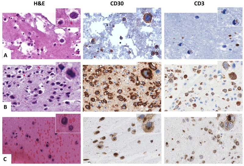
Fig 3. Immunophenotypic characterization of BI-ALCL cell blocks.
Representative cases are shown to underline the variability of CD3 expression in contrast to the consistent, intense and diffuse positivity for CD30 in BI-ALCL. In A CD3 was negative in almost all the tumor cells. In B CD3 staining was heterogeneous with the lymphomatous cells being CD3-negative, CD3-weakly positive and CD3-strongly positive. In C the majority of the neoplastic cells showed a faint CD3 expression (all original magnification x400).

