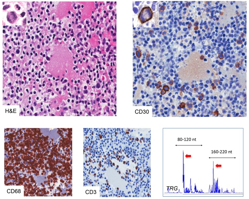
Fig 6. Morphological and molecular features of a CD30-rich reactive effusion.
The seroma was composed mainly by CD3+ T cells associated with scattered CD68+ macrophages and polymorphonucleates. Immunohistochemistry for CD30 stained a fraction of medium-large proliferating cells equal to 5% of the total cellularity (H&E, original magnification x200; mitotic figures in the inserts x400). TRG (Vg 9/11—Jg) clonality by Genescan fragment analysis showed a prominent peak (red arrows) within a polyclonal background in both the reference size ranges given by the BIOMED2 protocol (black arrows).

